
Big picture: The human brain = a 3-pound, 86-billion–neuron SaaS platform running 24/7. Evolution layered “legacy code” (hindbrain) under shiny “front-end” cortex. Know the hardware, know the psyc exam.
1. Structural Roadmap
| Evolutionary Layer | Key Parts | Elevator-Pitch Function | AP Hook |
|---|---|---|---|
| Hindbrain | Medulla – heartbeat / breathing Pons – sleep, facial expressions, bridge to cerebellum Reticular Formation – arousal filter Cerebellum – balance, implicit memory | Life-support & motor fine-tuning | Medulla damage = death; alcohol → cerebellar wobble |
| Midbrain | Superior/Inferior Colliculi – visual & auditory orienting Substantia Nigra – dopamine, movement | Sensory routing & movement trigger | Parkinson’s = substantia nigra DA loss |
| Forebrain (Diencephalon) | Thalamus – sensory switchboard (except smell) Hypothalamus – 4 F’s (fight, flee, feed, mate), controls pituitary Basal Ganglia – voluntary movement smoothing | Motivation & relay | Hypothalamus lesions alter appetite |
| Forebrain (Limbic) | Hippocampus – explicit memories Amygdala – emotion (fear/aggression) Septum – pleasure, relief | Emotion & memory hub | H.M. lost hippocampi → anterograde amnesia |
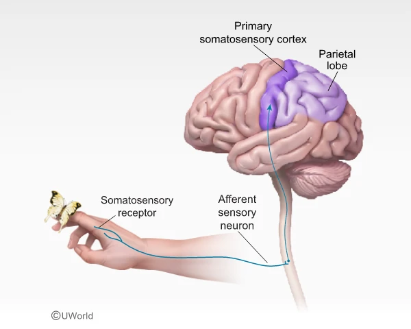
| Cerebral Cortex | Frontal lobe – motor cortex, prefrontal exec, Broca’s area (speech production) Parietal lobe – somatosensory cortex, spatial math Occipital lobe – vision (V1 etc.) Temporal lobe – hearing, Wernicke’s area (language comprehension) | High-level cognition & perception | Stroke in left frontal = Broca’s aphasia |
| Inter-Hemispheric | Corpus Callosum – fiber bridge | Lateralized info share | Split-brain studies (Sperry & Gazzaniga) |
2. Functional Hotspots
Motor Cortex (rear frontal): maps body parts → voluntary movement
Somatosensory Cortex (front parietal): touch & body position (homunculus)
Association Areas: vast “silent cortex” doing thinking, planning, face recog.
Prefrontal Cortex: working memory, inhibition, morality; last to mature (~25 yrs).
3. Brain Imaging Cheat Codes
| Method | Measures | Pros | Cons |
|---|---|---|---|
| EEG | Electrical activity (timing) | Millisecond precision; cheap | Poor spatial resolution |
| CT (CAT) | X-ray slices (structure) | Fast trauma scan | Radiation; low detail |
| MRI | Magnetic fields (structure) | High-res anatomy, no X-ray | Expensive; can’t show function |
| PET | Radioactive glucose (function) | Shows metabolism | Low res; radiation |
| fMRI | Blood-oxygen (function + structure) | Best of both worlds | Lag (~2 s), pricey |
4. Lateralization & Plasticity
Left-brain ≈ language, logic; Right-brain ≈ spatial, emotion, music.
Contralateral control: left cortex → right body, and vice-versa.
Plasticity: after injury, unused areas can re-wire; greatest in childhood.
10 AP-Style Multiple-Choice Questions
(Choose the best answer. After the table, detailed rationales follow.)
| # | Question | Options |
|---|---|---|
| 1 | Which brain area directly regulates hunger and body temperature? | A. Thalamus B. Hippocampus C. Hypothalamus D. Cerebellum E. Pons |
| 2 | Damage to the left motor cortex will most likely impair: | A. Speech comprehension B. Vision in right field C. Movement of the right hand D. Movement of the left hand E. Balance while standing |
| 3 | A patient understands speech but struggles to pronounce words. The lesion is probably in the: | A. Wernicke’s area B. Broca’s area C. Angular gyrus D. Somatosensory cortex E. Amygdala |
| 4 | The reticular formation is most involved in: | A. Coordinating fine motor movements B. Filtering incoming stimuli and arousal C. Regulating blood pressure D. Forming explicit memories E. Language syntax |
| 5 | Which technique would a researcher choose to watch real-time brain activation while a subject solves math problems, without using radioactive tracers? | A. EEG B. PET C. CT D. fMRI E. MRI |
| 6 | Split-brain patients shown an object in the left visual field can best: | A. Name the object aloud B. Point with right hand C. Point with left hand D. Write its name with right hand E. Describe its color verbally |
| 7 | Alcohol’s disruption of balance and coordination is tied to its depressant effect on the: | A. Cerebellum B. Hippocampus C. Medulla D. Basal ganglia E. Frontal pole |
| 8 | The limbic system structure that tags memories with emotional significance is the: | A. Thalamus B. Amygdala C. Pons D. Parietal lobe E. Substantia nigra |
| 9 | Which lobe integrates touch sensations with visual input for spatial judgment? | A. Temporal B. Occipital C. Parietal D. Frontal E. Insular |
| 10 | In long-term potentiation, repeated stimulation strengthens synaptic connections primarily in the: | A. Hypothalamus B. Cerebellum C. Hippocampus D. Medulla E. Corpus callosum |
Detailed Answers & Explanations
| # | Correct | Why It’s Right | Why Others Aren’t |
|---|---|---|---|
| 1 | C | Hypothalamus = homeostasis (hunger, temp, thirst). | Thalamus relays senses; cerebellum balance, etc. |
| 2 | D | Left motor cortex controls right body; damage impairs opposite side movement. | C flips laterality; others mismatch function. |
| 3 | B | Broca’s area = speech production; damage → non-fluent aphasia. | Wernicke’s is comprehension; others unrelated. |
| 4 | B | Reticular formation screens stimuli & maintains wakefulness. | A = cerebellum; C = medulla; D = hippocampus. |
| 5 | D | fMRI gives live functional maps sans radioactivity. | PET uses tracers; MRI gives structure only; EEG time-good but location-poor. |
| 6 | C | Info from left visual field → right hemisphere; right can’t speak but can guide left hand pointing. | Speech centers in left hemisphere, so naming fails. |
| 7 | A | Cerebellum handles coordination; alcohol disrupts its GABA signaling. | Others handle memory, autonomic, etc. |
| 8 | B | Amygdala = emotional processing/“fear tagger.” | Thalamus relay, etc. |
| 9 | C | Parietal integrates somatosensory + spatial mapping. | Temporal = auditory; occipital = vision, etc. |
| 10 | C | Hippocampus shows robust LTP → basis for memory formation. | Other areas not primary LTP showcase in AP syllabus. |
Free-Response Practice
FRQ #1 – Stroke Case (7 pts)
Question:
Mr. Lopez suffers a stroke that damages his right parietal lobe. He can still speak fluently but ignores objects on the left side of his visual field (left spatial neglect). Using psychological terminology, explain why each symptom occurs and predict one everyday activity that will now pose difficulty for Mr. Lopez.
High-Scoring Answer Sketch
Location (1 pt): Right parietal lobe integrates spatial & sensory info from left visual field/body.
Neglect (2 pts): Damage disrupts contralateral spatial awareness → left spatial neglect (he ignores left side).
Speech spared (1 pt): Language centers (Broca, Wernicke) reside mainly in left hemisphere, unaffected.
Everyday impact (2 pts): He may shave only the right side of his face or eat food from only the right half of a plate.
Compensation (1 pt): Occupational therapy can leverage brain plasticity to retrain attention.
FRQ #2 – Comparing Imaging Methods (7 pts)
Question:
A cognitive neuroscientist wants to study decision-making speed while minimizing participant risk. Compare EEG, PET, and fMRI in terms of (a) what each measures, (b) temporal versus spatial resolution, and (c) practical/ethical considerations. Recommend the best method and justify.
High-Scoring Answer Outline
| Method | Measures (1 pt each) | Resolution (1 pt each) | Practical/Ethical (1 pt each) |
|---|---|---|---|
| EEG | Surface electrical potentials | Excellent temporal (ms), poor spatial | Inexpensive, non-invasive, but can’t pinpoint deep structures |
| PET | Radio-tagged glucose metabolism | Decent spatial, poor temporal (min) | Radioactive tracer → ethical concern, costly |
| fMRI | Blood-oxygen-level changes (BOLD) | Good spatial (mm) & moderate temporal (~2 s) | No radiation; loud/expensive, motion sensitive |
Recommendation (1 pt): fMRI balances spatial detail with acceptable temporal lag and avoids radiation, suiting real-time decision tasks ethically.